
Translate this page into:
Role of Negative Pressure Wound Therapy in Diabetic Foot Ulcers: A Case Report
Corresponding author: Aditya Chebrolu MBBS Konaseema Institute of Medical Sciences & Research Foundation, Amalapuram, Andhra Pradesh, India Email ID - adityakumar1215@gmail.com
-
Received: ,
Accepted: ,
Abstract
Diabetic foot ulcers (DFUs) are a leading cause of hospitalization among diabetic patients and represent a significant global complication of diabetes mellitus. Annually, approximately 18.6 million individuals develop DFUs, which are a precursor to 80% of lower extremity amputations in diabetic patients and are associated with higher mortality rates. Effective management of DFUs requires a multidiscipli-nary approach involving podiatrists, infectious disease specialists, and vascular surgeons, which has been shown to reduce major amputation rates compared to standard care. Among current treatment modalities, Negative Pressure Wound Therapy (NPWT) is a widely recommended and non-invasive technique that applies controlled negative pressure to accelerate the healing of complex diabetic foot wounds. NPWT has been demonstrated to be both safe and effective in promoting faster wound closure and granulation tissue formation. This report discusses the case of a 49-year-old female with a chronic DFU on the plantar surface of her left foot, which was successfully managed with NPWT. Following its application, significant wound size reduction and the early formation of healthy granulation tissue were observed. The case underscores the vital role of NPWT in enhancing the healing process of DFUs, offering a valuable treatment option for reducing complications and improving patient outcomes.
Keywords
Diabetic foot ulcer
Negative pressure wound therapy
Debridement
Ray amputation
Proteus
Klebsiella
Meropenem

INTRODUCTION
Diabetic foot ulcers (DFUs) are a significant complication of diabetes mellitus, contributing to morbidity and an increased risk of lower extremity amputation. DFUs are often associated with neuropathy and peripheral vascular disease, which impair wound healing and infection control.1 A multidisciplinary approach, involving surgical debridement, antibiotic therapy, and advanced wound management techniques, is essential for improving patient outcomes.2 Negative Pressure Wound Therapy (NPWT) has emerged as an effective modality in promoting wound healing by reducing edema, increasing perfusion, and facilitating granulation tissue formation.3 This case report discusses the management of a 49-year-old female patient with a chronic DFU on the plantar surface of her left foot, treated successfully with NPWT, emphasizing its importance in reducing wound size and enhancing healing.
CASE REPORT
A 49-year-old female with a 20-year history of diabetes mellitus, hypertension, and coronary artery disease (CAD) presented to the casualty department with fever, chills, and a chronic oozing wound on the plantar surface of her left foot. The patient had been on insulin therapy for diabetes and aspirin for CAD. One month prior to admission, the patient sustained a cut on the plantar side of her left little toe after stepping on small rocks during a family event. Due to diabetic neuropathy, the injury went unnoticed, and she did not seek medical attention promptly. The wound was managed with frequent dressings for one month before her admission.
On general examination, the patient was febrile with an axillary temperature of 101.5°F. Physical examination revealed a 5x4x3 cm ulcer on the plantar surface of the left foot with purulent discharge [Figure-1]. An X-ray of the left foot showed normal tarsal bone morphology, but destructive changes were observed in the surrounding soft tissues.
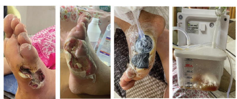
- Chronic diabetic foot ulcer (DFU) on the plantar surface of the left foot before and after surgical debridement, negative pressure wound therapy (NPWT) was applied using a vacuum-assisted closure (VAC) system.
Based on clinical, radiologic, and laboratory findings, surgical intervention was deemed necessary. During the procedure, necrotic tissue was identified in both the dermal and subdermal layers, along with purulent involvement of the plantar fascia. Debridement and necrectomy were performed, followed by ray amputation of the fifth toe. Intraoperative swabs were taken for both aerobic and anaerobic cultures. The patient was started on intravenous meropenem (1 g twice daily) in the hospital ward. On postoperative day 4, the microbiological cultures tested positive for Proteus and Klebsiella species. Antibiotic therapy with meropenem was continued based on the antibiogram results. Daily wound dressings with normal saline, hydrogen peroxide, and papain ointment were performed.
On postoperative day 10, after assessing a reduction in wound size, negative pressure wound therapy (NPWT) was initiated at a constant pressure of -125 mmHg. NPWT was applied for 5 days, promoting further wound healing. On postoperative day 14, the NPWT device was removed, revealing a significant reduction in wound size and excellent granulation tissue formation. After 3 days of subsequent wound dressings, a split-thickness skin graft was placed over the wound to facilitate closure. The patient responded well to the treatment, and follow-up care was planned to ensure full recovery.
DISCUSSION
The Vacuum-Assisted Closure (VAC) system is an advanced technology in wound management that has proven effective in treating complex wounds, including diabetic foot ulcers (DFUs).1,2 DFUs are notoriously challenging due to poor vascularization, neuropathy, and increased risk of infection. VAC therapy, also known as Negative Pressure Wound Therapy (NPWT), provides a controlled, subatmospheric pressure environment that promotes faster healing. The system comprises four key components: a filler substance or sponge placed inside the wound, a semipermeable dressing to isolate the wound from the environment, a vacuum system, and a connecting tube to transfer pressure to the wound.3,4
Effective VAC therapy requires careful preparation, beginning with radical debridement to remove necrotic or devitalized tissue. The first VAC dressing is typically applied in an operating room under sterile conditions by trained medical personnel. Hemostasis is critical before applying standard negative pressure of 125 mm Hg, although lower pressures (75 mm Hg) may be used if excessive bleeding persists. Kalstostat dressings can be applied temporarily for cases with continuous bleeding before transitioning to VAC therapy.4,5
The foam used in the VAC system is particularly effective, as its open-pore structure allows uniform distribution of pressure across the wound, optimizing fluid drainage and wound contraction. Proper placement of the foam is crucial to ensure that it covers the entire wound surface. The foam, tubing, and surrounding healthy tissue must be sealed with an adhesive drape to maintain an airtight environment.3,5
NPWT works through multiple mechanisms, including macro- and micro-deformation of the wound bed, fluid removal, and stabilization of the wound environment. These actions stimulate granulation tissue formation, angiogenesis, and neurogenesis, thereby enhancing wound healing. Studies have demonstrated that VAC therapy reduces wound size, increases blood flow, and stabilizes the wound environment, making it particularly useful for DFUs. The intermittent suction mode is preferred over continuous suction, as it promotes better tissue oxygenation and granulation.3,5
While highly effective, VAC therapy does have complications, such as pain, bleeding, and foam retention. A loss of suction is a common mechanical issue, often caused by an inadequate seal or improper placement of the suction drain. Managing these challenges requires careful attention to detail, ensuring proper application of dressings and regular monitoring. Additionally, clinicians must be vigilant in preventing dehydration and monitoring for hypersensitivity reactions or further tissue damage. Overall, VAC therapy provides a valuable tool in the management of DFUs, improving wound healing rates and reducing the risk of amputation when applied with precision and proper care.3,5
CONCLUSION
In conclusion, NPWT has proven to be a highly effective treatment for diabetic foot ulcers, significantly enhancing wound healing and infection control. Its benefits include improved drainage, increased granulation tissue formation, and reduced bacterial load, which accelerate the healing of chronic and deep wounds common in diabetes. However, patient selection is critical, as improper use can lead to severe complications. Future research should focus on refining NPWT protocols and exploring combinations with other advanced therapies to improve outcomes. Importantly, diabetic patients should regularly check their feet for wounds and keep them moisturized to prevent complications, as early intervention is key to better management of diabetic foot ulcers.
END NOTE
Author Information
Aditya Chebrolu, MBBS, Konaseema Institute of Medical Sciences & Research Foundation, Amalapuram, Andhra Pradesh, India Haripriya Thikkireddy, MBBS, Konaseema Institute of Medical Sciences & Research Foundation, Amalapuram, Andhra Pradesh, India
Conflict of Interest
None declared
References
- Diabetic Foot Ulcers: A Review. JAMA. 2023;330(1):62-75.
- [CrossRef] [PubMed] [PubMed Central] [Google Scholar]
- Diabetic foot ulcer: A comprehensive review of pathophysiology and management modalities. World J Clin Cases. 2023;11(8):1684-1693.
- [CrossRef] [PubMed] [PubMed Central] [Google Scholar]
- Consensus on the application of negative pressure wound therapy of diabetic foot wounds. Burns Trauma. 2021;9:tkab018.
- [CrossRef] [PubMed] [PubMed Central] [Google Scholar]
- Diabetic foot ulcers: Classification, risk factors and management. World J Diabetes. 2022;13(12):1049-1065.
- [CrossRef] [PubMed] [PubMed Central] [Google Scholar]
- Negative pressure wound therapy for treating foot wounds in people with diabetes mellitus. Cochrane Database Syst Rev. 2018;10(10):CD010318.
- [CrossRef] [PubMed] [PubMed Central] [Google Scholar]


